General ECG Information
Holter Monitoring
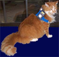
Holter monitoring is a noninvasive method of assessing the heart's rhythm and rate. It is essentially an electrocardiogram that is recorded while the animal is wearing a recorder. This is usually for 24 hours but sometimes up to 48 hours or even a week. This allows for an analysis of the rhythm and rate of the heart throughout the monitoring period with activities documented in a diary by the owner which we provide. Your veterinarian may recommend this diagnostic procedure for the following reasons:
- An arrhythmia is suspected in your pet
- Your pet exhibits signs of weakness, dizziness, lethargy, excessive panting and agitation, or even collapse or fainting episodes
- To monitor drug therapy and effects on the heart rate and rhythm
- To monitor programming effects and detect malfunctions if your pet has an implanted pacemaker
This is the most thorough an accurate test to document any arrhythmias and slow or high heart rates throughout the monitoring period.
Cardiac Event Monitoring
As one of a very few companies that provide weekly cardiac event monitoring and have done so for over 20 years, we excel at what we do.
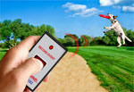
A cardiac event monitor is a battery powered portable device that you control to record primarily your pets hearts electrical activity when they are having symptoms. Sometimes your veterinarian may want to document the effects of a new or altered dose of your pets medication over the period of a week.
There are 2 types of event monitors, one has a memory (this one is worn continuously) and the other is placed on the chest area when a symptom occurs or to do a simple recording. Both monitors record only when you press the button to record. Unlike a Holter which will record the 24/48 hours in it's entirety, not missing a beat.
These monitors are used to document slow or fast heart rates, early or extra beats and even "missed" beats. These conditions can case symptoms of dizziness, collapsing, exercise intolerance or lethargy.
The recordings are stored and sent to us through your phone (preferably a land line which has a clearer connection for better viewing of the ECG complexes). We then instruct you on how to send it to us and we then are able to view and analyze the ECG data. Your veterinarian will then be sent the results if abnormal or at the end of the monitoring period if normal.
Why do you need this test?
Regular 12 lead ECGs done in your doctors office allow the doctor to see your heart's electrical activity for less than 1 minute. Abnormal heart rhythms come and go so this may not be enough time for your doctor to "catch" the abnormality. An event or Holter monitor allows your doctor a greater period of time to detect any abnormalities.
Are there any risks to using the Holter or Event Monitor such as my pet getting shocked?
No, are no risks to using either type of monitor. Both monitors use AA or AAA batteries so no chance of getting shocked if they bite through the wires.
What dogs or cats get arrhythmias?
All breeds and ages of dogs and cats can get arrhythmias. Some specific arrhythmias are identified in specific breeds. The cause and the treatment vary widely depending on the diagnosis.
Ventricular Arrhythmias
Boxers, bulldogs, German shepherds
-
A common disease identified in boxers is arrhythmogenic right ventricular cardiomyopathy or ARVC. This is commonly called "Boxer Cardiomyopathy". The arrhythmia seen in these dogs is primarily from the right ventricle, but they may also come from other locations in the heart. Bulldogs also get a variation of this disorder. These ventricular arrhythmias may occur in rapid succession and this is called ventricular tachycardia. When ventricular tachycardia occurs it may lead to a decrease in blood flow to the body. When the perfusion to the brain decreases enough dogs may collapse. This arrhythmias may degenerate into ventricular fibrillation which is a fatal abnormal rhythm. Therefore, some dogs with ventricular arrhythmias must be treated with antiarrhythmics (e.g. sotalol). Most often to determine if this is required electrocardiograms are recorded and 24-hour electrocardiograms are monitored (Holter monitoring). These same tests are used to monitor the response to treatment. In addition to treating the arrhythmias associated with ARVC, dogs need to have other diagnostics to understand the extent of the structural and functional problems in addition to the electrical disorder of the arrhythmia.
German shepherds have an inherited ventricular arrhythmia that affects young dogs between 3 and 24 months of age. Some dogs die suddenly of these arrhythmias most commonly between 5 and 9 months of age. A diagnosis usually requires a Holter monitoring period to catch the dangerous arrhythmia. After 24 months of age the arrhythmias can disappear and the dogs are no longer at risk, although when used for breeding they have affected offspring when mated to a dog that has the genetic background of risk.
Atrial Fibrillation
Great Danes, Doberman Pinschers, Newfoundlands, Irish Wolfhounds, Boxers and other large breeds
- Atrial fibrillation may be associated with underlying cardiomyopathy that has resulted in poor heart function and enlargement of the heart or it may occur with when the heart is structurally and functionally within normal range. The diagnosis is made from an electrocardiogram. However, most importantly, for the best care, 24-hour ambulatory electrocardiographic monitoring (Holter monitoring) is required to fully understand the rhythm disturbance. Atrial fibrillation most commonly causes the heart to beat too fast. In dogs, cardioversion may be successful in returning the heart to a normal rhythm, but often the rhythm returns to atrial fibrillation in the dog. Consequently, treatment for atrial fibrillation involves the use of drugs to slow the rate. Such drugs include dilitiazem, atenolol and/or digoxin. It is vital in the management of this arrhythmia that follow-up Holter monitoring be used to optimize treatment with the correct dose. Follow-up echocardiography and radiography also are important to determine the need for other medications.
Sick Sinus Syndrome
West Highland White Terriers, Dachshunds, Miniature Schnauzers, Boxers, Cocker spaniels
-
Sick sinus syndrome is characterized by a heart rhythm whereby the sinus node (which normally initiates the beating heart) does not discharge an impulse to trigger the heart to contract. As a result the heart literally stops beating. If the heart stops for more than 8 seconds then the dog will collapse/faint. Sometimes the heart will have another part of the heart initiate a beat to rescue the heart from complete arrest. Most of the time the sinus node will eventually start up again to do its job but the dog has a rhythm with many long pauses. Some dogs with sick sinus syndrome have a more constant sinus bradycardia (too slow) because the sinus node has a low firing rate. Other dogs with sick sinus syndrome will have periods of excessive tachycardia (rapid rate) in addition to the pauses or bradycardia. When a dog has clinical signs of sick sinus syndrome it is almost always required that a pacemaker be implanted. The implantation of a pacemaker is today a common procedure in dogs. Veterinary cardiologists who are experienced in the implantation of pacemakers and the programming of these pacemakers can best insure the best treatment for afflicted dogs. The response to treatment is usually very good. In dogs that also have the tachycardia this is treated with medication(s) if it does not subside after pacing.
Heart Block
Any breed, may affect cats too
- Normally the heart beat is initiated by the part of the heart called the sinus node. The impulse conducts through the atria and to the junctional point between the atrial and the ventricles. The junction is known as the atrioventricular node or AV node, in short. Here, the conduction is slowed allowing time for the atria to fill the ventricles with blood before the ventricles are triggered to contract. In dogs and cats with heart block the conduction to the ventricles is not just slowed, but eliminated. This means that the ventricles are not told to contract. Most animals will have secondary pacemakers in the conduction system of the ventricles trigger the heart to beat when the conduction through the AV node is stopped. This saves the animal. These secondary pacemakers do not discharge at the same rate as the primary natural pacemaker of the sinus node. Instead, they go slowly. Sometimes dangerously slow. This causes the animals to be weak and even collapse. Heart failure can actually develop. The majority of animals with this type arrhythmia must be treated with a pacemaker. Before a pacemaker is implanted the animals are evaluated thoroughly by echocardiography and other testing (e.g. troponin I concentrations) to determine other underlying problems.
Myocarditis
Any breed but often medium to larger dogs
- Some dogs with ventricular arrhythmias or heart block may have underlying inflammation of the heart known as myocarditis. In these situations monitoring of the arrhythmia and other tests (e.g. echocardiography, troponin I, C-reactive protein) are required. Often the cause of the myocarditis cannot be identified, but this disease quite often must be handled carefully because often dogs die suddenly when affected. Treatment includes antiarrhythmic medications and anti-inflammatory drugs such as corticosteroids.
Canine Dilated Cardiomyopathy (DCM)
CAUSE:
Canine dilated cardiomyopathy (DCM) is a primary disease of cardiac muscle that results in a decreased ability of the heart to generate pressure to pump blood through the vascular system. The definitive cause of canine DCM is the subject of debate, although a number of factors including nutritional, infectious, and genetic predisposition have been implicated. The fact that canine DCM occurs at a higher incidence in specific breeds suggests a heritable genetic component to this disease, although it is likely that it’s etiology is multifactorial. Breeds predisposed to DCM include the Doberman Pinscher, the Great Dane, the Boxer, and the Cocker Spaniel. Dietary carnitine deficiency may play a role in some cases of Boxer DCM, and taurine responsive DCM has been identified in Cocker Spaniels.
CLINICAL SIGNS:
DCM is characterized by dilation of the ventricles with ventricular wall thinning. In many cases, dilation of all four chambers of the heart is seen. The ability of the heart to serve as a pump is diminished, and clinical signs of DCM occur secondary to either decreased delivery of oxygenated blood to the body (lethargy, weakness, weight loss, collapse), or to congestion of blood in the lungs (coughing, increased respiratory rate and/or effort, abdominal distention) or both. Cardiac dilation, decreased oxygen supply, and increased oxygen demand secondary to elevated heart rate and ventricular wall stress may predispose to the development of cardiac arrhythmias arising in either the atria (atrial fibrillation, supraventricular tachycardia) or in the ventricles (ventricular premature complexes, ventricular tachycardia). Arrhythmias may predispose affected dogs to sudden death.
DIAGNOSIS:
DCM is diagnosed by echocardiography, which demonstrates the chamber dilation and indices of decreased pump function characteristic of the disease. Thoracic radiography is useful to evaluate pulmonary (lung) tissue and vessels, and may show evidence of fluid accumulation in the lungs (pulmonary edema) or around the lungs (pleural effusion). Electrocardiography may be used to characterize heart rhythm and to rule out arrhythmias; and in some cases, a 24 hour electrocardiogram (Holter monitor) may be recommended to more accurately characterize cardiac rhythm.
TREATMENT:
Treatment of DCM is directed at improving systolic (pump) function of the heart, dilating the peripheral blood vessels to decrease ventricular workload, eliminating pulmonary congestion if present, and controlling heart rate and cardiac arrhythmias if present. These treatment goals are addressed by the administration of cardiac medications, which may be delivered by injection in emergent situations, or orally in patients that are more stable.
PROGNOSIS:
Canine DCM can be a devastating disease, and the prognosis for dogs with DCM is variable depending upon breed and status at presentation. The prognosis for Doberman Pinschers with DCM, for example, is less favorable than in other breeds, while DCM in Cocker Spaniels may be relatively slowly progressive. Patients that present in congestive heart failure generally have a worse prognosis than those that are not in congestive heart failure at presentation. Irrespective of this, medical therapy may provide significant improvement in lifespan and quality of life in affected dogs.
Feline Hypertrophic Cardiomyopathy (HCM)
The most commonly diagnosed cardiac disease in cats.
Hypertrophic cardiomyopathy (HCM) is Characterized by regional or diffuse thickening of the walls of the ventricle (the primary "pump" muscle of the heart), HCM has been diagnosed in cats as young as 4 months old and as old as 16 years old. Although the definitive cause of feline HCM has not been identified, its prevalence within certain breeds (i.e. Maine Coon cats, Ragdolls) has prompted speculation that at least some forms of HCM are genetic in origin. The finding of mutations in an important cardiac protein called myosin binding protein C in affected lines of Maine Coon and Ragdoll cats supports a heritable, genetic component of HCM in these breeds.
Thickening of the walls of the ventricle is associated with a decreased ventricular chamber volume and abnormal ventricular relaxation (diastolic function) in cats with HCM. Since the amount of blood pumped by the heart per minute (cardiac output) is the product of the amount of blood ejected per contraction (stroke volume) and the heart rate in beats per minute, this decreased chamber volume (and subsequent stroke volume) results in an increased heart rate (tachycardia) as a reflex mechanism to maintain cardiac output and blood pressure. Although this reflex increase in heart rate may maintain normal blood pressure in the short term, it is associated with an increased consumption of oxygen by the heart muscle, to the extent that oxygen demand may exceed supply. This scenario may result in an energy starved heart muscle, with subsequent heart cell death and worsening function. Another consequence of an increased heart rate is that the ventricle has less time to fill between contractions, further diminishing stroke volume and promoting a vicious cycle of reflex tachycardia, decreased time for ventricular filling, and so on. This decreased left ventricular filling also promotes stasis of blood in the left atrium (chamber just before the left ventricle), which ultimately contributes to the development of clinical signs (see below).
CLINICAL SIGNS:
Many cats with HCM present without overt signs of illness. In other cases, signs of congestive heart failure including labored or rapid breathing, open-mouth breathing, and lethargy are evident. These signs occur when fluid accumulates in lung tissue (pulmonary edema) or around the lungs (pleural effusion) secondary to elevation of left atrial pressure. A potentially devastating cclud of HCM is thromboembolism. Thromboembolism refers to the development of a clot in the heart (promoted by left atrial enlargement), with ejection of the clot to the systemic circulation. When the clot lodges in the peripheral circulation, it may obstruct blood flow to the region of the heart supplied by the blocked vessel. The site of thromboembolism most commonly observed in cats with HCM is the distal aorta (termed a saddle thrombus), and clinical signs of hind limb paralysis and acute pain in the hind limbs may be observed. Thromboembolism is a poor prognostic indicator in cats with HCM.
DIAGNOSIS:
HCM is diagnosed by echocardiography, which shows the characteristic thickening of the left ventricular walls and decreased chamber volume of the left ventricle. Evaluation of the left atrium for dilation and/or the presence of a thrombus are also achieved using this modality. Since hyperthyroidism and hypertension may also cause left ventricular thickening, these diseases must be ruled out prior to arriving at a diagnosis of HCM. Thoracic radiography may be useful to evaluate pulmonary (lung) status and to rule out pleural effusion. Electrocardiography may be useful to characterize heart rate and to rule out cardiac arrhythmias.
TREATMENT:
Treatment goals for feline HCM include controlling heart rate, alleviating pulmonary congestion, removing pleural fluid (if present), and decreasing the likelihood of thromboembolism. These goals are addressed by the administration of medications that may be delivered by injection in emergent situations or orally in more stable patients. Some drugs (i.e. nitroglycerine) may be applied to the skin for absorption.
PROGNOSIS:
The prognosis for cats with HCM is quite variable. Cats without clinical signs may survive for years, although the disease is most commonly progressive. Poor prognostic indicators include the presence of congestive heart failure, thromboembolism, and hypothermia (low body temperature). In spite of the fact that HCM may decrease the life span of affected cats (sometimes significantly so), medical therapy can improve the quality of life and longevity of cats affected with this common disease.
Press Release - March 17,2023
Pet Cardiology Receives 2022 Best of El Dorado Hills Award
Pet Cardiology has been selected for the 2022 Best of El Dorado Hills Award in the Veterinarian category by the El Dorado Hills AwardProgram.
Each year, the El Dorado Hills Award Program identifies companies that we believe have achieved exceptional marketing success in their local community and business category.These are local companies that enhance the positive image of small business through serviceto their customers and our community. These exceptional companies help make the ElDorado Hills area a great place to live, work and play.
Various sources of information were gathered and analyzed to choose the winners in each category. The El Dorado Hills Award Program focuses on quality, not quantity. Winnersare determined based on the information gathered both internally by the El Dorado HillsAward Program and data provided by third parties.
About El Dorado Hills Award Program
The El Dorado Hills Award Program is an annual awards program honoring the achievementsand accomplishments of local businesses throughout the El Dorado Hills area. Recognition isgiven to those companies that have shown the ability to use their best practices andimplemented programs to generate competitive advantages and long-term value.
The El Dorado Hills Award Program was established to recognize the best of local businessesin our community. Our organization works exclusively with local business owners, tradegroups, professional associations and other business advertising and marketing groups. Our mission is to recognize the small business community's contributions to the U.S. economy.
Testimonials
"I am a veterinarian who was in sudden need of a Holter monitor for my own pet. After I had foolishly waited several days for a competing service to get back to me, I called Forest at Pet Cardiology and he got the monitor to me by noon the next day (across the country!). The service was fantastic, 24hour help not only for vets but also for owners, both of which I used.
Middle of the night calls were dealt with beautifully. Unfortunately my pet Emma died suddenly while wearing the Holter. The compassion showed to me by the entire company demonstrated a true dedication to animals and their owners. The report was thorough, detailed and sent to me promptly, and the price was more than reasonable, especially considering the excellent service. I will be using this service for my clients from now on and will be recommending it highly to my peers. Thanks Forest and Pet Cardiology!"
--Colleen Cosker, DVM Connecticut
"I just wanted to let you know how pleased I was with your service. Everyone was so helpful! I received the monitor by Fedex two days after my initial contact. Return packaging was included, and I had telephone confirmation of the results within 24 hours of your receipt of the returned monitor. Actually, only a week passed from the time I first called to my receipt of the telephoned results! Thank you so much!"
Connie Haywood
Boxers at Wit's End
Davidsonville, Maryland 21035
"In the six years that I have worked with Global Telemedicine/Pet Cardiology, they have provided prompt, reliable, and courteous service. They are available to our hospital and our clients 24 hours a day. The staff is always friendly and helpful and they provide very competitive pricing for their services."
Carl D. Sammarco, BVSc, MRCVS, Diplomate ACVIM (Cardiology)
Red Bank Veterinary Hospital www.rbvh.net
"I have used your service for years, speedy, accurate and have never been disappointed"
Stacy L. Hosking, DVM, DACVIM (Cardiology) Nevada
www.cardiovet.net
"The company was very efficient and the staff was great"
A. Legender DVM Tennessee


